A tiny speck of dust in reality might not be home to thousands of microscopic creatures living in a fully built city complete with a mayor, but when put under a microscope it can reveal some still fascinating details. Today we round up 10 of the best microscopic images ever captured.
No 10. Diatom rainbows
Captured through polarizing light filters, this image is courtesy of a retired British microscopist Michael Stringer and it grabbed the top position in 2008 Nikon Small World Photomicrography Competition. The image shows sinewy filaments within squirming microscopic diatoms.
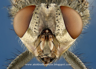
No 9. Muscoid fly
Remember this guy? Yeah, you guessed it; it is a common house fly, magnified 6.25 times. Captured by Chrales B. Krebs for 2005 Nikon Small World Photomicrography.
No 8. Arabidopsis thaliana (thale cress) anther
This image of a male sex organ of flowering plant comes in at number 8. Magnified 20 times, this image was captured by Estonian scientist Heiti Paves of the Tallin University of technology. This image has been chosen for its scientific and artistic qualities for a record 2000 entries.
No 7. Double transgenic mouse embryo
Magnified 17 times the original size, this image illustrates a late stage developing mouse embryo, specifically after 18.5 days. It was captured by Gloria Kwon. This image was a runner-up in 2007 Nikon Small World Photomicrography competition.
No 6. Human tongue taste bud
Showing one of the 10 thousand taste buds in the human tongue, this image was captured using Scanning Electron Microscope, also known as SEM by David Gregory & Debbie Marshall.
No 5. Beetle dancing on a pin
Ever wondered how many angels could dance on the head of a pin? Well, keep wondering because I don’t have the answer also, but what I do have to give you is, this beautiful image of a tiny leaf beetle dancing, not dancing really, but standing over the head of a pin. This image is a 40x magnification of the original thing, and everything including the beetle’s rainbow colors, is all natural.
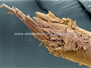
No 4. Split hair
Coming right out of the Head & Shoulders commercial is this image of a split hair. This image by Liz Hirst, Welcome Images, should motivate you enough to regularly get your hair trimmed or to use a good conditioner to avoid this kind of ugly picture.
No 3. Red blood cells
This beautiful image of red blood cells which we humans carry in amounts of trillions, comes in at number 2. In women there are about 4 to 5 million RBCs per micro liter (cubic millimeter) of blood and about 5 to 6 million in men.
No 2. Influenza virus neuraminidase
This visually beautiful image shows crystals of influenza virus neuraminidase separated from terns and is a 14x magnification. This image by Julie Macklin and Dr. Graeme Lave was a runner-up in the 1987 Nikon Small World Photomicrography competition.
No 1. Individual Gold atoms
While not as visually appealing as other images in this list, this image symbolizes a special achievement. This image was produced by TEAM 0.5, world’s most powerful transmission electron microscope in 2008, though I am not aware of its current status. If someone from our readers want to enlighten us on this, then please do in the comments section. The image clearly distinguishes between the individual atoms which wasn’t previously possible. To give you an idea of how big an achievement this is, the distance between the individual atoms you can see is 2.3 angstroms and 1 angstrom is equal to 10^(-10) meters.


0 Komentar:
Posting Komentar